
Incubator Accessories

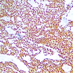
Diagnostic Biosystems OCT 3/4 OCT-3/4 (OCT4/6847R)
OCT-3/4 (also known as POU5F1) is a transcription factor that has been recognized as fundamental in the maintenance of pluripotency in embryonic stem cells and primordial germ cells. It has been proposed as a useful marker for germ cell tumors (GCT) that exhibit features of pluripotentiality (seminoma/dysgerminoma, germinoma and embryonal carcinoma. OCT-3/4 immunostaining has been shown to be sensitive and specific for GCT, whether in primary gonadal or extragonadal sites or in metastatic lesions.

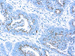
Diagnostic Biosystems CASPASE 3/CPP32 Caspase 3/CPP32
This antibody reacts with the active form of caspase 3 (17 kD protein). Caspase 3 is a member of the interleukin-1 beta-converting enzyme family and is thought to be associated with the induction of apoptosis. Caspase 3 is synthesized as an inactive 32 kD proenzyme and is processed during apoptosis generating two subunits of 17 kD and 12 kD. Caspase 3 stains the epithelial cells of skin, renal proximal tubules, and collecting ducts.
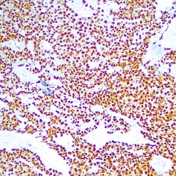
Diagnostic Biosystems OCT 3/4 OCT-3/4 (OCT4/6847R)
OCT-3/4 (also known as POU5F1) is a transcription factor that has been recognized as fundamental in the maintenance of pluripotency in embryonic stem cells and primordial germ cells. It has been proposed as a useful marker for germ cell tumors (GCT) that exhibit features of pluripotentiality (seminoma/dysgerminoma, germinoma and embryonal carcinoma. OCT-3/4 immunostaining has been shown to be sensitive and specific for GCT, whether in primary gonadal or extragonadal sites or in metastatic lesions.
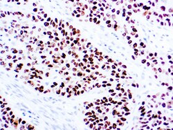
Diagnostic Biosystems P40 p40
p40 is a relatively unknown antibody that recognizes ΔNp63-a p63 isoform suggested to be highly specific for squamous/basal cells. In a recent study, p40 is equivalent to p63 in sensitivity for squamous cell carcinoma, but it is markedly superior to p63 in specificity1, which eliminates a potential pitfall of misinterpreting a p63-positive adenocarcinoma or unsuspected lymphoma as squamous cell carcinoma. These findings strongly support the routine use of p40 in place of p63 for the diagnosis of pulmonary squamous cell carcinoma.
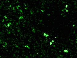
Diagnostic Biosystems C1Q-FITC C1q Complement-FITC
This antibody reacts with the human C1q complement. Traces of contaminating antibodies have been removed by solid phase absorption with human plasma proteins. This antibody is well suited for tissue section staining but may also be used for other applications.
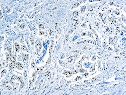
Diagnostic Biosystems CYCLIN D1 Cyclin D1 (SP4)
Cyclin D1 or PRAD-1 or bcl-1 is one of the key cell cycle regulators, and functions in association with cdk4 and/or cdk6 by phosphorylating the Rb protein. It is a putative proto-oncogene overexpressed in a wide variety of human neoplasms including mantle cell lymphomas (MCL).
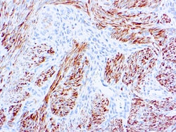
Diagnostic Biosystems MOUSE AND RABBIT LINK Desmin (D33)
Cytoskeletal intermediate filaments (IFs) constitute a diverse group of proteins that are expressed in a highly tissue-specific manner. IFs are constructed from two-chain α-helical coiled-coil molecules arranged on an imperfect helical lattice, and have been widely used as markers for distinguishing individual cell types within a tissue and identifying the origins of metastatic tumors. Vimentin is an IF general marker of cells originating in the mesenchyme. Vimentin and Desmin, a related class III IF, are both expressed during skeletal muscle development. Desmin, a 469 amino acid protein found near the Z line in sarcomeres, is expressed more frequently in adult differentiated state tissues. Anti-desmin detects cells of normal smooth, skeletal, and cardiac muscles. Antibody reacts with leiomyomas, leiomyosarcoma, rhabdomyomas, rhabdomyosarcoma, and perivascular cells of glomus tumors of the skin.
Diagnostic Biosystems HistoZyme
HistoZyme is a proteolytic enzyme solution that results in superior staining over traditionally used enzyme pretreatments, such as pepsin and trypsin. Moreover, HistoZyme requires only a brief incubation at room temperature. Performing antigen recovery with HistoZyme, will allow you to dilute the primary antibody 5 to 10 fold further and achieve optimal staining.\n\nFormalin fixation of tissues induces protein cross links that helps in maintaining the cellular morphology by inactivating the digestive enzymes and preserving the cytoskeleton. Fixation stops tissue autolysis, preserves tissue structures, and immobilizes antigens. However, antigens undergo chemical alteration of their primary, secondary and tertiary structures during fixation.

Diagnostic Biosystems FLUOROMOUNT Fluoromount™
Fluoromount™ is an innovative, aqueous-based mounting medium designed especially for the permanent preservation of fluorescent specimens. Fluoromount™is to be used with tissue sections stained with a multitude of fluorescent dyes. This product is compatible with FITC, phycoerythrin, phycocyanin, and allophycocyanin. It can also be used with other fluorescent markers such as Texas Red and rhodamine.Mounting tissue specimens stained with fluorescent dyes.

Diagnostic Biosystems BLUE HEMATOXYLIN KIT Blue Hematoxylin Kit
DBS's Blue Hematoxylin Kit is a histological staining reagent suitable for visualization of nuclei in tissue sections. Blue Hematoxylin is a blue nuclear counterstain. Bluing Reagent is used following Counterstaining both designed for optimal performance when used in immunohistochemistry.\n\nDBS’s Blue Hematoxylin is intended for use in the histologic demonstration of nuclear staining. This staining technique is used to make the critical distinction between a normal nucleus and an abnormal one. When used as a counterstain in immunohstochemical procedures, Blue Hematoxylin is compatible with most commonly used chromogens including DBS HRP-Yellow, HRP-Red, HRP-Green, HRP-Black, and DAB. When applied to tissue sections, these will change the hue of hematoxylin from purple to blue It replaces the strong alkaline rinses or long tap water washes which may cause loss of cells or tissue sections.

Fritsch Milling And Sizing Inc Stacking Ring for 125-150Ml Premium Line
Stacking ring for all grinding bowls for Planetary Mill PULVERISETTE 5 premium line 125-150 ml volume.
Essential, if 2 grinding bowls 125-150 ml volume per grinding bowl holder are used.

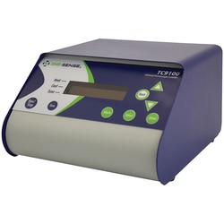
Cole-Parmer Digi-Sense TC9100 Benchtop Temperature Controller for Thermocouples; 230 VAC
The benchtop single-loop temperature controllers are designed to regulate a user-defined set point temperature. The microprocessor control provides heating/cooling tolerances for demanding applications found in laboratory or industrial environments. Temperature scale selectable to read in F, C, K (Kelvin), or R (Rankine). An auto-tuning function provides automatic configuration of the PID parameters with minimum user configuration required. Nonvolatile memory stores all user-defined information. A convenient output receptacle, located on the back of the units, allows for direct plug-in of heaters and other devices. All models accept eight thermocouple types (J, K,N,R,S,T B, and E). Safety features include a loop-break alarm, overtemperature and timer cutoffs, separate fuses for instrument and output receptacle, and audible/visible out-of-range alarms.
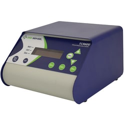
Cole-Parmer Digi-Sense TC9600 Benchtop Temperature Controller with USB Output, for Thermocouple, RTD, or Thermistors; 230 VAC
The benchtop single-loop temperature controllers are designed to regulate a user-defined set point temperature. The microprocessor control provides heating/cooling tolerances for demanding applications found in laboratory or industrial environments. Temperature scale selectable to read in °F, °C, K (Kelvin), or °R (Rankine). An auto-tuning function provides automatic configuration of the PID parameters with minimum user configuration required. Nonvolatile memory stores all user-defined information. A convenient output receptacle, located on the back of the units, allows for direct plug-in of heaters and other devices. All models accept eight thermocouple types (J, K,N,R,S,T B, and E). Safety features include a loop-break alarm, overtemperature and timer cutoffs, separate fuses for instrument and output receptacle, and audible/visible out-of-range alarms.
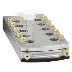
Cole-Parmer ATS20100 Reflux/Inerting Unit for Integrity 10
Keep your reaction station running smoothly with these parts and accessories.
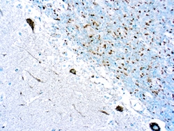
Diagnostic Biosystems PAN-TRK Pan-TRK
Neurotrophic tyrosine kinase receptor (NTRK) is a family of 3 proto-oncogenes including NTRK1, NTRK2, and NTRK3 which encode Trk A, Trk B, and Trk C proteins, respectively. The latter are involved in biological processes such as neuronal survival, differentiation, and plasticity under physiological circumstances. In various cancers, oncogenic fusionsinvolving the kinase domain of NTRK1, 2, or 3 are present. Pan-Trk IHC is a time- and tissue-efficient screen for NTRK fusions, particularly in driver-negative advanced malignancies and potential cases of secretory carcinoma and congenital fibrosarcoma. Pan-Trk IHC can help determine whether translation occurs for novel NTRK rearrangements.